BY HEATHER SMITH THOMAS
Arthroscopy is a common procedure for diagnosis and treatment of problems inside a joint. The word arthroscopy comes from two Greek words: arthro, meaning joint; and scope, meaning look.
Arthroscopic surgery is a minimally invasive surgical procedure in which examination and treatment is performed using an arthroscope—an endoscope inserted into the joint through a small incision. Arthroscopy (in the joint), laparoscopy (into the abdomen), thoracoscopy (in the chest), etc. are minimally invasive compared to traditional surgeries.
Dr. Marcos Perez, a boarded surgeon at Peterson Smith Equine Hospital + Complete Care in Ocala, Florida does many of these surgeries in horses—mainly joint repair and colic surgeries.
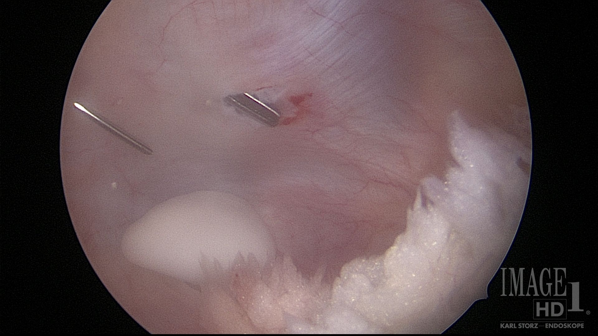
“Arthroscopy was first used in horses in 1974 and developed in the 1980’s so that when we have to go inside a joint, we can do the smallest possible incision—with less disruption of tissues,” Perez said. “There is less damage to the joint and joint capsule—hence less inflammation from the surgery. This can also decrease the risk of infection; there is not such a big opening to the outside.”

Arthroscopic procedure – Photo Courtesy: Dr. Marcos Perez
Typically, four-millimeter cannulas are used and slightly larger incisions are used to insert those, but nearly everything is done with two small incisions—one for the scope and one for the instruments. Occasionally the surgeon has to make more than one instrument portal, but it is still a very small incision. If there’s a bone fragment larger than a chip fracture, the length of the small instrument incision is enlarged to be able to pull out the fragments.
Arthroscopy has been a huge technological advantage for diagnosis and treatment for joint problems.
“We also do laparoscopies to check inside the abdomen, but the main use in horses is for minimally invasive procedures inside the joints,” Perez said.
“We use this to guide procedures; when we repair fractures that go through a joint, we can make sure the bone pieces are perfectly aligned, with no defects in the bones when we put them back together in the joint. We can also remove little fragments, or clean up ragged parts from injured tendons, or bone spurs that might cause adhesions.”
With many OCD (osteochondrosis dissecans) problems in horses, most of the joint is fine at the time the surgery is done; the defect or injury is small. This kind of surgery in young horses is more of a preventative, to keep bad things from happening with fragments or bone chips that can move and become lodged, creating more cartilage damage. There are also many things that can be done with tendons and tendon sheaths. The horse has uniquely important flexor tendons that sometimes suffer injury inside the tendon sheath, which can be diagnosed and treated by tenoscopy.
“We don’t do as many of these, because there are fewer pathologies in tendon sheaths than in joints. Repairing a tendon might be a bit more tricky using tenoscopy but we can also shave off all the broken fibers and clean it up for better healing.”
A tiny camera is utilized to visualize what’s inside the joint.
Arthrocopy – Floating fragment loose in the stifle – Photo Courtesy: Dr. Marcos Perez
“It is usually four millimeters (0.16 inches) to 25 millimeters (slightly less than an inch). Arthroscopy is the main diagnostic tool to assess cartilage damage because cartilage does not show up on radiographs. You can sometimes see a tiny bit with ultrasound or MRI or contrast CT but what you see with the arthroscopy camera is like what you’d see with your own eyes and understand what is wrong with the joint.”
Stifle injuries can also be treated with arthroscopy.
“We do a lot of meniscus surgeries, but the meniscus in the horse is difficult to treat because there is not much space between those two bones,” Perez said. “We can only clean the surface of the meniscus when it is damaged, so this type of injury may not have a good prognosis. In humans, this is easier [with knee surgery].”
“We do about 200 arthroscopies a year at Peterson Smith. There are other equine hospitals in this area that do this, so there are probably about 600 horses here each year that are diagnosed and treated with arthroscopy,” he says. It’s a common procedure.
There are very few complications after this type of surgery.
“In terms of cosmetic results, most of the time you can’t see where it was performed, which is good/desirable for owners.”
It’s also a big advantage for racehorses and sport horses that must be perfectly sound for their jobs.

Fragment removed from a sesamoid bone – Photo Courtesy: Dr. Marcos Perez
“We do a lot of racehorse arthroscopy, especially in babies that have OCD lesions and chip fracture fragments,” Perez said. “Those clean up very well and improve the prognosis of the joint so they can race longer. Also, once they fall free in the joint, we can also remove any fragments that are moving around, damaging the cartilage.”
The techniques have continually improved, and today there are new developments such as 3D arthroscopy.
“You can see the depth of the joint and not just a flat screen. I don’t think it makes much difference for a surgeon who has been well trained and doing these surgeries for many years, but for the new generation of surgeons, just learning to do this, it may help them visualize things and triangulate better because they don’t yet have as much experience,” Perez said.
Laparoscopy has also evolved and improved.
“The equipment has improved and image quality is much better. The cameras and images now are all HD (high definition) and 4K. You need light to see inside the abdomen or the joint, and those lights—Xenon or LED–have improved and now create less heat, with more power or lumens.”
Light is very helpful inside the joint or abdomen.
“When you are looking around in a big cavity that is very dark, you need a big light but it has to go through a small, minimally invasive space,” Perez said.
“Whenever we look inside a joint we use sterile fluid to distend and lavage the joint fluid, and whenever we are in the abdomen we use gas—usually CO2—to inflate the area so we can look around easier,” he said.
“This creates more space within the joint, or the abdomen. Whenever it tends to close down so we can’t see, we distend the cavity with fluid or gas so those walls open up more and we can see better,” Perez said. “Those systems have been improved and have different settings—so we don’t over-pressurize the joints or squeeze the lungs; now we can control this so we don’t do any damage with too much pressure.”

Laparoscopy is utilized a lot, not only for colic diagnosis and surgeries but also for checking ovaries and remove tumors in ovaries.
“We can do this with the horse standing and sedated. I also use this for cryptorchid surgeries. Male horses with a testicle that has not descended into the scrotum—retained within the abdomen—can be more easily treated with laparoscopy. We can go into the abdomen with a scope and find it quicker; we can remove the testicle in about 20 minutes instead of searching for two or three hours trying to find it.”
This is much easier on the horse, and the person doing it.
There are courses in arthroscopic surgery, and ongoing research.
“Colorado State University has been the lead in arthroscopic research and they usually have one basic and one advanced course each year for residents or surgeons who want to improve. For horses, arthroscopy is the gold standard for joint surgery.”
Return to the March 13 issue of Wire to Wire